
The LCPTM Distal Fibula Plates has created a locking compression plate system that combines traditional plating methods with locking screw technology.
Titanium and stainless steel are the material options for the plates.
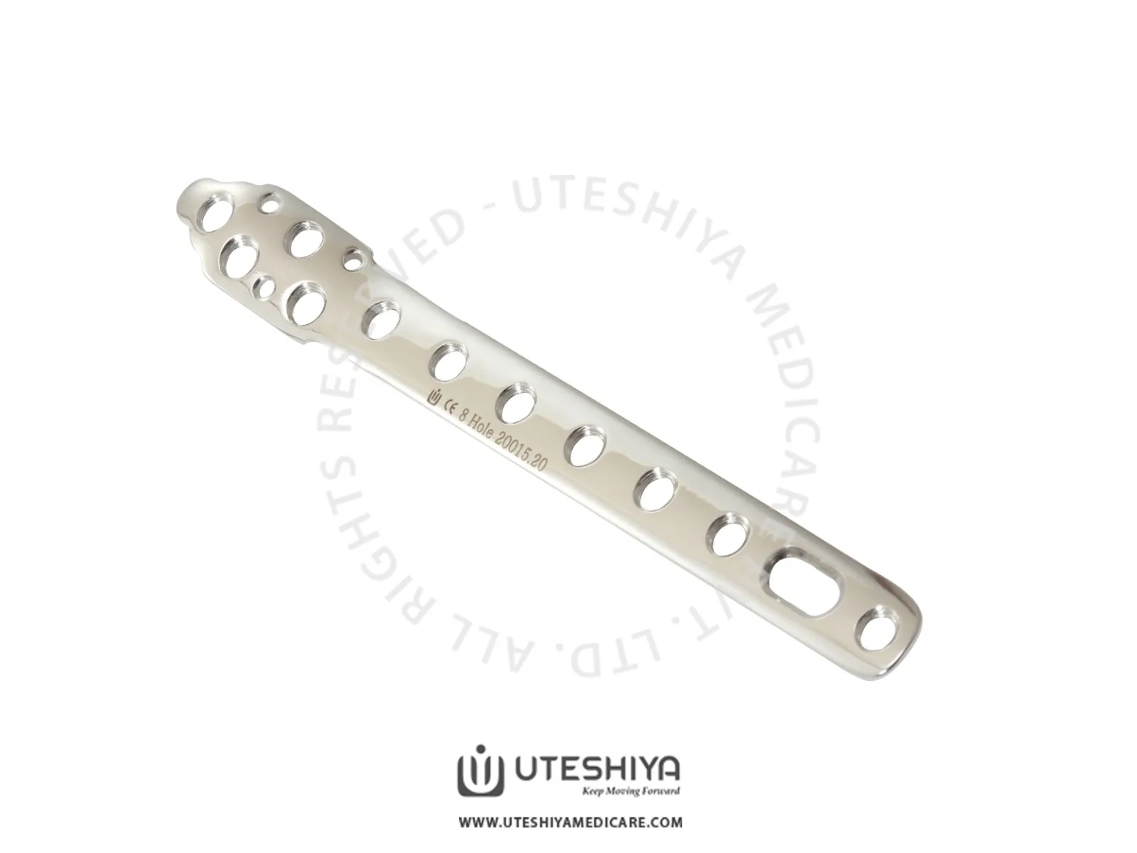
Along the fibular shaft and distally, the plates have anatomical features. The LCP plate shaft’s combi-holes are a combination of a locking screw hole and a dynamic compression unit (DCU) hole.
Combi-holes provide axial compression and locking capabilities throughout the whole plate shaft, giving you more versatility. You may temporarily secure the plate to the distal fibula and determine its placement relative to the bone by using Kirschner wires (up to 2.0 mm) through the holes in the plate.
By using locking screws, one may manufacture a fixed-angle structure using conventional AO plating methods. Instead of resisting patient weight by plate-to-bone compression, these screws work like multiple small, angled blade plates.
Describe the anatomy of the Distal Fibula
One of the lower leg bones, the distal fibula, is located outside the leg and is smaller than the other. It is also the name of the bone. It is a very important factor in ankle stability, especially when it comes to responsibilities that require weight-bearing.
It is possible for this area of the body to sustain a fracture as a result of trauma or repeated stress, and it may be challenging to treat because of the complex structure of the bone and its position that it is located in the ankle joint.
Understanding LCP Distal Fibula Plate
Orthopedic implants like the LCP (Locking Compression Plate) Distal Fibula Plate help heal fractures and other damage to the lower leg’s smaller fibula bone. This plate is an integral aspect of Synthes’s LCPTM system, which integrates locking screw technology with conventional plating techniques.
Important Features
- Surgeons can choose between titanium and stainless steel when it comes to the LCP Distal Fibula Plate so they may meet the individual demands of their patients.
- The anatomical design of the plates is such a tight match to the distal fibula’s form that they provide an ideal fit and maximum stability.
- Along the plate shaft, you’ll find “combi-holes,” which are a mix of locking screw holes and DCU holes. Combi-holes provide a versatile solution for fracture repair, allowing for both axial compression and locking capabilities.
- Kirschner wires (K-wires) inserted through the plate’s holes provide a temporary fixation solution that helps determine the plate’s eventual location by securing it to the bone.
- Locking screws like a set of tiny, angled blade plates, these screws form a structure with a fixed angle. As a result, the fracture is better able to recover and remains stable.
Surgical Technique in 7 steps
Applying for the 3.5mm LCP Distal Fibula Plate is all things considered, a complex process that calls for expert skills. On the other hand, when done properly, it may provide patients with distal fibula fractures with strong fixation and the best possible results.
To make sure the LCP Distal Fibula Plate is properly positioned and stable, the surgical approach in question requires several important stages. The general procedure of the surgery is as follows:
Step: 1
Surgeons look at their patients’ X-rays and CT scans before surgery to determine the fracture pattern and figure out how to approach the procedure. Based on the severity and location of the fracture, the surgeon will decide what size and kind of plate is necessary.
Step: 2
Depending on the surgeon’s discretion and the precise pattern of the fracture, the patient is either put in a supine or lateral position on the operating table.
Step: 3
Making an incision along the bone’s natural way allows the surgeon to expose the distal fibula. Careful dissection of the soft tissues reveals the bone and fracture location.
Step: 4
Reducing a fracture involves careful manipulation and alignment of the fracture pieces to return the distal fibula to its standard structure. A pair of reduction forceps or similar tools could be necessary for this work.
Step: 5
The LCP Distal Fibula Plate, which is shaped to conform to the bone, is attached to the side of the fibula. A stable fixation is achieved by positioning the plate to cover the fracture location.
Step: 6
The next step is to put locking screws into the inside of the bone using the combi-holes in the plate. Stabilizing the fracture and encouraging bone healing, these screws form a fixed-angle construct. It may be necessary to insert more screws to provide a secure hold.
Step: 7
After the plate and screws are inserted, the incision is layered closed. The surgical site is covered after applying a sterile dressing.
Postoperative Care and Rehabilitation
Proper healing and excellent results after 3.5mm LCP Distal Fibula Plate surgery depend on postoperative care and therapy. As a whole, these are the standards for postoperative care and recovery:
- After surgery, the patient’s ankle and lower leg are immobilized in a cast or brace. This helps to preserve the surgical site and promotes healthy bone repair.
- When it comes to weight bearing, the surgeon will give you particular instructions. After certain types of leg surgeries, patients can put all their weight on the treated leg right away; however, others will need the help of crutches or a walker for a while.
- It is possible to give pain medication to aid with the management of pain after surgery. Patients should take the medicine exactly as prescribed and notify their healthcare workers if they have any severe pain.
- After surgery, the patient must take special care to keep the incision dry and clean. Regarding matters like showers and dressing changes, they have to strictly adhere to the surgeon’s advice.
- An important component of any effective rehabilitation plan is physical therapy. With the help of a physical therapist, the patient will be able to increase their ankle joint strength, flexibility, and function.
- Mild stretching, weight training, and balancing drills are all possible workouts.
- Patients should schedule frequent follow-up appointments with their surgeon after surgery. The surgeon will look at the patient’s recovery and overall health during these checkups. It is possible to use X-rays to determine where the surface of the plate and screws are.
- How long it takes for a patient to go back to their regular routine after a fracture depends on their specific condition and how quickly they heal. As far as activity limitations and a gradual return to activities are concerned, patients should listen to their surgeon’s advice.
- Infection, slowed healing, or hardware failure are all possible problems that patients should be aware of. They need to be quick to inform their doctor if they have any strange symptoms.
What are the complications and solutions
| Complication | Solution |
| Infection | Administer appropriate antibiotics |
| Surgical debridement if necessary | |
| Hardware Failure | Revision surgery to replace or reposition hardware |
| Use of stronger or additional fixation devices | |
| Malunion | Surgical correction to realign the bone |
| Physical therapy to restore function | |
| Nerve Damage | Surgical exploration and repair if necessary |
| Physical therapy to regain function and sensation | |
| Joint Stiffness | Physical therapy and range of motion exercises |
| Hardware Irritation | Removal of hardware if symptoms persist |
| Modification of weight-bearing activities |
Comparison with Other Fixation Techniques
When compared to more conventional methods of fixing fractures of the distal fibula, the LCP Distal Fibula Plate provides a number of benefits that were not before available. A comparison may be made as follows:
- When compared to conventional plates and screws, this connection method is more stable.
- Increased anatomical compatibility, which may result in a lower chance of malunion or nonunion
- Combination holes that allow for compression and locking, allowing for more flexibility in fracture fixation
- a minimally invasive procedure that has the ability to reduce disruption of soft tissue
- This allows for early weight-bearing, which in turn promotes bone repair and reduces the development of problems.
Future Developments and Innovations
Improvements in patient outcomes and reductions in the risk of complications are anticipated to be the focus of future developments with the LCP Distal Fibula Plate since the field of orthopedic surgery is continually expanding. The following are examples of possible advances and inventions in the future.
- Plates with improved design that provide a better anatomical fit and maintain stability
- The use of biodegradable implants would eliminate the need for surgical removal.
- The use of 3D printing technology to create individualized implants for each patient, as well as improvements in imaging methods that allow for more precise planning and navigation
- Implants with bioactive properties that enable bone repair and integrate more quickly
Wrapping It Up
When it comes to treating distal fibula fractures, the LCP Distal Fibula Plate is really innovative. It outperforms conventional fixation techniques in terms of stability and results because of its anatomical design, adaptability, and locking screw technology. Postoperative care and rehabilitation are as important as the surgical approach for a speedy recovery. It is believed that this innovative treatment method will be even more successful in the future because of changes in orthopedic surgery, such as biodegradable implants and better plate design.