 About 17% of all fractures and about 1.5-2.5% of all visits to emergency rooms are due to distal radius fractures (DRFs), making them a prominent acute occurrence in traumatology.
Fractures of the metacarpal bones (10.5% of all fractures), the proximal femur (8.9% of all fractures), and the finger phalanges (11.2%) rank highest in terms of frequency in the general population.
Recent investigations have shown that over half of distal radius fractures (DRFs) are linked to ulnar styloid fractures and that nearly all distal forearm fractures, 98.3%, are radius fractures, which can be connected with ulna fractures.
What is an external fixator?
A stabilizing structure called an external fixator is used to keep the fractured bones in the correct alignment. By making tiny cuts in the muscle and skin, an external fixator inserts metal screws or pins into the bone. A bar outside the skin holds the screws and pins.
Having an external fixator could make even the most routine tasks appear more difficult at times. If you’re finding it difficult to tackle any of these chores, these helpful suggestions may do the trick.
Traditional Approaches to Fracture Management
Many different ways have been used for treating fractures in the past, and these are all part of the traditional fracture therapy strategy. Usually, there are six methods that are commonly used and very popular in fracture management. Factors such as the kind and level of the fracture, patient characteristics, and the treating physician’s preferences and competence determine the use of each of these conventional methods, which each have its own indications, advantages, and limits.
1. Casting Method
Plaster or fiberglass casts immobilize the injured region while they heal by supporting and aligning the fractured bones.
2. Splinting treatment
Treatment of injuries often involves the use of splints, which can be either stiff or flexible, to immobilize and support the affected region.
3. Traction process
Using pulleys and weights to apply stress to the injured limb to straighten bones and minimize fractures before or during surgical procedures is known as traction.
4. Bracing process
Bracing is the process of stabilizing and supporting the wounded region while allowing regulated mobility. Orthoses and braces are often composed of metal, plastic, or other materials.
5. Closed Reduction method
Closed reduction is a non-surgical method of repositioning broken bones, often done with the patient under either local or general anesthetic.
6. Open Reduction treatment
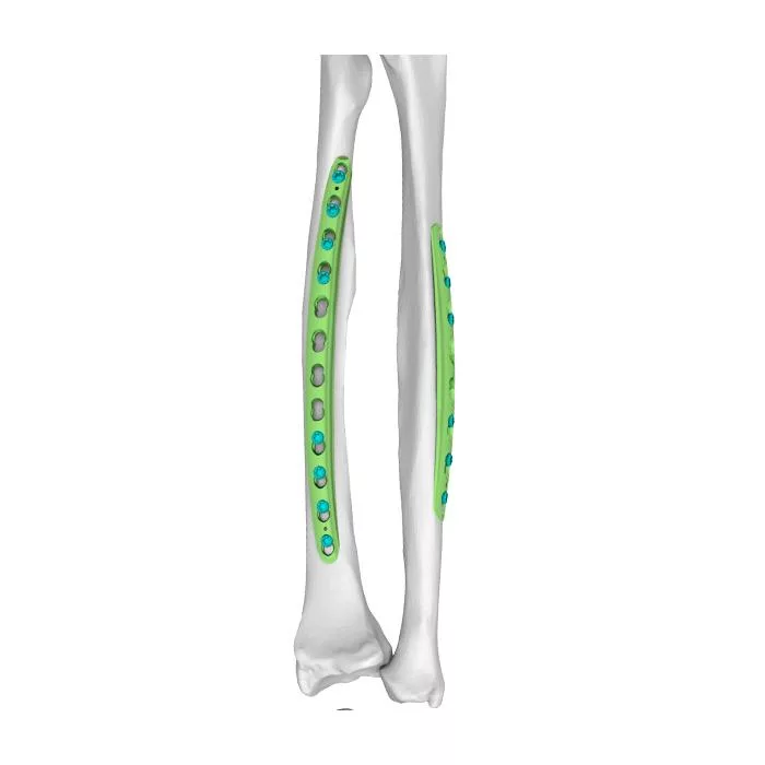
An open reduction is a surgical procedure that exposes broken bones so that screws, plates, or other fixation devices can straighten and stabilize them.
Factors such as the kind and level of the fracture, patient characteristics, and the treating physician’s preferences and competence determine the use of each of these conventional methods, which have indications, advantages, and limits.
Evolution of External Fixation
During the first part of the nineteenth century, the main concerns surrounding surgery were the possibility of dangerous infections and related pain. This meant that non-operative treatment was the gold standard for fractures and dislocations.
The three main inventions that shaped the evolution of surgical therapy for fractures were anesthesia (1846), antisepsis (1865), and X-rays (1895). It is often believed that Malgaigne was the first to employ external fixation (1843). It would be a stretch to call his equipment an external fixation. Typically, the first “real fixator” was Lambotte’s 1902 exterior fixation device. It all began in 1897 when Clayton Parkhill introduced his “bone clamp” to the American market.
Technological Innovations
Orthopedic devices that stabilize high-energy and complicated fractures of the extremities, known as external fixators, are difficult and costly. Despite rapid technological advancement, these devices’ mechanical fracture stabilization aims have not altered in decades.
Orthopedics may benefit from 3D printing external fixation devices. We analyze and synthesize the literature on 3D-printed external fixation devices for orthopedic trauma fractures.
Methods
The PRISMA (Preferred Reporting Items for Systematic Review and Meta-Analysis) procedures were followed for this paper with slight modifications. Two independent reviewers filtered the search results using 3D printing and external fracture fixation inclusion and exclusion criteria.
There is a great deal of variation in the external fixator designs and testing methodologies found in the existing research on this particular topic.
The use of 3D printing in this subspecialty of orthopedic surgery has been the subject of a few publications in the peer-reviewed literature. Several preliminary clinical case studies have shown encouraging outcomes from using 3D-printed external fixation designs. However, bigger trials with standardized testing and reporting are needed.
Future Trends for Health Care
New technologies are transforming fracture therapy with the promise of improved accuracy, efficiency, and patient comfort.
There will likely be a greater emphasis on individualized patient care, better results, and more sophisticated procedures in the future of external fixation.
The future of external fixation for fracture healing looks bright, despite the fact that there are a lot of challenges to overcome.
A more effective and patient-friendly fracture treatment setting is possible through embracing change and new ideas.
Challenges and Considerations
In veterinary orthopedics, external fixation is a good way to stabilize many fractures, although it isn’t perfect. Following the rules for fixator selection and application is the best approach to reduce the possibility of issues. Problems with the bone-pin contact, fixator failure to sustain reduction and fracture healing are major difficulties that could affect the effectiveness of fixing the fracture.
Pintract drainage, inadequate limb usage, and neurovascular damage are minor problems that might cause discomfort or annoyance. Most problems may be dealt with if they are identified quickly, their causes identified, and suitable treatment administered.
Here are some additional challenges and considerations of external fixation in distal ulna and radius fractures.
| Challenges | Considerations |
| Fracture complexity requires precise fixation. | Careful patient selection is crucial. |
| The device limits mobility, impacting daily life. | Scheduled follow-ups track healing progress. |
| Accurate placement of pins/screws is critical. | Post-op care instructions are essential. |
| Complications risk necessitates vigilant care. | Patient education enhances adherence to care. |
| Potential soft tissue irritation from hardware. | Rehabilitation plans aid in recovery. |
Wrapping It Up
An external fixator is a tried-and-true method that orthopedic surgeons have used for decades. By carefully following the instructions outlined in this article, it has the potential to be a reliable method for treating distal radius fractures.