What is a PLIF Cage?
Posterior lumbar interbody fusion (PLIF) is a surgery done from behind the lumbar spine and tries to fuse two levels of the spine.
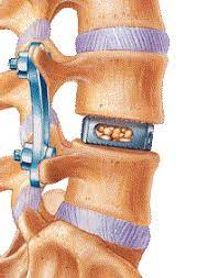
It is created for insertion between adjacent vertebrae in the lower back in order to promote fusion and restore spinal column stability. The cage is usually made of a biocompatible material, such as titanium or polyetheretherketone (PEEK). It has a hollow, cage-like structure with an enormous surface area and open spaces.
A scaffold for new bone development is created by placing bone graft material within the cage to facilitate fusion. The posterior lumbar interbody fusion cage also helps keep the discs’ height, realign the spine, and relieve pressure on the nerves, all of which are important to the success of the fusion operation as a whole.
Advantages of PLIF Cages
-> PLIF cages stabilize bone graft material, promoting spinal fusion.
-> PLIF cages straighten and stabilize the spine, lowering pain and improving spinal function.
-> The cage restores disc height between vertebrae, which reduces nerve pressure and improves spinal biomechanics.
-> Enhanced load-bearing capacity. Titanium or PEEK PLIF cages can handle spinal column forces.
-> Compared to open procedures, PLIF cages can be introduced through small incisions, decreasing tissue damage, blood loss, and recovery time.
-> PLIF cages can be tailored to each patient’s anatomy.
-> PLIF cages can relieve nerve pressure while facilitating interbody fusion.
-> The posterior lumbar interbody fusion cage becomes part of the spinal column after fusion, providing long-term support.
Considerations when Choosing a PLIF Cage
PLIF cage selection requires numerous considerations. The cage’s compatibility with the patient’s physiology comes first. Titanium or PEEK should be biocompatible and safe.
To meet the patient’s anatomy and surgical technique, the cage’s size and design should be carefully considered during cage selection. To improve fusion rates and stability, evaluate the cage’s surface area, porosity, and lordotic angle.
The surgeon should consider the patient’s spinal degeneration, alignment, and biomechanics before cage selection. To avoid difficulties, cage insertion surgical technique and instrumentation must be assessed.
Finally, the surgeon should consider PLIF cage availability, cost-effectiveness, and long-term clinical outcomes. Selecting the best PLIF cage for spinal fusion requires surgeon-patient collaboration.
Surgical Technique for PLIF Cage Placement
Spine surgeons do posterior lumbar interbody fusion surgery in hospitals or outpatient centers.
The procedure takes 2 to 6 hours, depending on the surgeon’s skill, the patient’s health, any co-existing disorders such as spondylolisthesis, and the number of spinal levels.
Open PLIF surgery typically involves these steps:
The spine is accessed through a 3-inch to 6-inch midline incision in the back. Both sides and many lamina levels are dissected from the left and right lower rear muscles (erector spinae). The spinous process, lamina, facet joints, and transverse processes are exposed for each spinal level.
A PLIF at the L4-L5 spinal section would expose the L4 spinous procedure, lamina, facet joints, and transverse processes of the vertebrae.
The spinous process of the affected level is removed, accompanied by laminectomy to decompress and see the midline thecal sac, dura, and releasing nerve roots on both sides.
Once enough space has been identified, the facet joints can be undercut (trimmed) to clear the area around the nerve roots.
The nerve roots are gently retracted midline to reach the posterior disc annulus. The annulus and disc material are cut.
Prepare the disc plates for fusing. Repeat on the other side of the disc. The side spaces are for interbody implants and bone graft material.
Instrumentation includes putting two interbody spacers and a bone graft into the space between the vertebrae.
Screws are placed bilaterally above and below the joined vertebrae, then connected with short metal rods to stabilize the vertebral bodies during fusion. The bone grows between vertebral bodies.
Minimizing Complications and Optimizing Recovery
-> Thorough preoperative planning: Assessing the patient’s medical history, imaging exams, and health can uncover risk factors and maximize surgical planning.
-> Experienced surgical team: Choosing an experienced PLIF surgeon can prevent complications and enhance surgical outcomes.
-> Proper patient selection: Assessing patients’ spinal status, overall health, and lifestyle before PLIF surgery helps reduce difficulties.
-> Navigation systems: Advanced imaging and navigation technology can help surgeons precisely place the PLIF cage and reduce problems.
Real-Life Success Stories and Patient Testimonials
-> People thinking about getting PLIF surgery might find it helpful to read about the experiences of patients who have had the process and had good results.
-> PLIF surgery testimonials might help prospective patients comprehend the potential benefits and gain confidence in their decision-making.
-> Sharing experiences about successful fusion, reduced pain medication use, and return to desired activities might demonstrate PLIF surgery’s sound effects on patients.
Future Developments and Innovations in PLIF Cages
-> Advanced biomaterials
New cage materials with improved biocompatibility, strength, and bone fusion may result from biomaterial research.
-> 3D-printed cages
Patient-specific PLIF cages, adapted to individual anatomy, can improve surgical results and patient satisfaction.
-> Biologics and growth factors
PLIF cages with biologics and growth factors may promote faster and more substantial bone fusion, accelerating recovery and minimizing non-union risk.
-> Navigation and robotics
Surgical navigation systems and robotics may allow more precise and minimally invasive PLIF cage placement, minimizing surgical complications and increasing patient outcomes.
Intraoperative imaging and real-time navigation can help surgeons visualize and guide PLIF surgery, enhancing accuracy and results.
Conclusion
Posterior lumbar interbody fusion (PLIF) cages have changed spinal surgery, improving the treatment of spinal diseases. Surgeons use PLIF cages to achieve effective spinal fusion and relieve symptoms by creating a stable environment for fusion and facilitating alignment. With continued developments and a patient-centered approach, PLIF cages will continue to bring hope, alleviation, and regained functionality to spinal disease patients.